Internal Anatomy & Physiology
General
The internal anatomy of sea anemones is quite unique compared to the rest of Cnidaria. To investigate the internal anatomy of C. polypus, specimens were sectioned along the sagittal plane to get an entire view of the specimen. Anatomical and micro-anatomical structures that have been used to classify Calliactis sp. are: a circular or ovoid base; a smooth column; the presence of cinclides; basitrichs located in the column; and 6 pairs of perfect sterile mesenteries. (Gusmão, 2010)
Figure 1 shows an illustration of the internal and external anatomy of the sea anemone. Important anatomical structures have been labelled in order to give and overall view of the anatomy of C. polypus.

|
| Figure 1: Labelled illustration of the internal and external anatomy of a sea anemone. (Shick, 1991) |
Figure 2 shows the sagittal section of a C. polypus specimen after it has been stained with hematoxylin and eosin. These stains were used to bring out all of the different tissues present in the anemone in order to view them under a microscope successfully.

|
Figure 2: Composite image of the sagittal section of C. polypus stained with hematoxylin and eosin. Important anatomical structures have been labelled.
|
Figure 3 is a cross section of the column of a sea anemone. It has been included to give an internal reference for Figure 1 in the Physical Description.
Musculature
In polyps, the muscles are distributed in opposing sheets of circular and longitudinal fibres. Usually the epidermal musculature is longitudinal and the gastrodermal is circular, both of which are smooth muscle. (Ruppert, 2004)
Nervous System
The nervous system of Calliactis polypus consists of superficial sensory neurons that monitor the environment, interneurons that join the sensory receptors to motornuerons, which activate effectors such as muscles or cnidocytes. The interconnected neurons form a pair of complex, two-dimensional nerve nets, located at the base of the epidermis and gastrodermis respectively. They are joined by neurons that bridge the mesoglea. (Ruppert, 2004)
Nematocysts
One of the defining characteristics of phylum Cnidaria are the nematocysts, or cnidae. Nematocysts are a type of cell, which aids in prey capture and defence against predators. Each nematocyst is made up of a capsule that contains a coiled tubule, usually with barbs. Toxin is also often contained within the nematocyst. Nematocysts have previously been described as independent of neural pathways (Pantin, 1942), however there is now evidence for two-cell and three-cell neural pathways between different types of nematocysts (Westfall, 2004).
25-30 morphological types of nematocysts exist within Cnidaria (Kass-Simon and Scappaticci, 2002, Fautin, 2009). Nematocysts are ejected as a response to stimuli via the innervation of a trigger hair, usually located on the cell itself. The nematocysts then fires and the tubule turns inside out and pierces or sticks to the predator or prey. Once the nematocyst has fired, the cell can not be used again, therefore a mixture of chemical and mechanical stimulation is required when setting off the trigger hair in order to save energy (Kass-Simon and Scappaticci, 2002). Like 60% of examined anemone genera, C. polypus has 3 types of nematocysts (Mariscal, 1974). These include basitrichs, microbasic p-mastigophores, and spirocysts (Fautin, 2013). Basitrichs are located all over C. polypus (Fautin, 2013, Gusmão and Daly, 2010) and have been found to function as both a prey capture mechanism and shell adhesive. (Mariscal, 1974). A distribution of the different nematocysts is located in Table 1.
|
|
Basitrichs
|
Microbasic p-mastigophores
|
Spirocysts
|
|
Acontia
|
X
|
|
|
|
Actinopharynx
|
X
|
|
|
|
Column
|
X
|
X
|
|
|
Mesenterial Filaments
|
X
|
X
|
|
|
Tentacles
|
X
|
|
X
|
Table 1: Adapted from (Fautin, 2013)
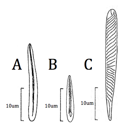
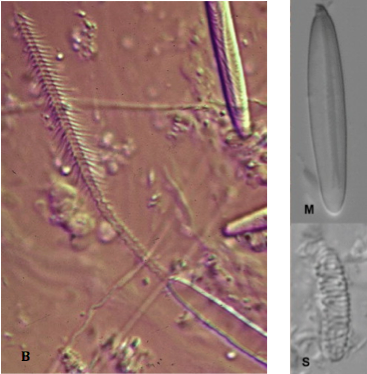
Figure 4 and 5 show the different types of nematocysts present in C. polypus. Photo B of Figure 5 shows the fired basitrich and the barbs associated with the tubule that is expelled.
|
| Figure 4: The three types of unfired nematocyst found in C. polypus and their relative and actual sizes. A)Basitrich; B)Microbasic p-mastigophores; C)Spirocysts. (Fautin, 2013) |
|
Figure 5: Fired and unfired nematocysts specific to C. polypus under a microscope (Fautin, 2009). B) Basitrich, M) Microbasic p-mastigophores, S) Spirocysts. For an idea on the scale of the images refer Fig 4 (as one was not provided in Fautin's paper).
|
(Mackie, 2002) stated, “They have enabled the group to achieve enormous success as predators with little of the investment in elaborate sensory and morphological specialization that characterizes most predators. Thus, cnidarians have prevailed despite their exceedingly simple basic body plan.”
|